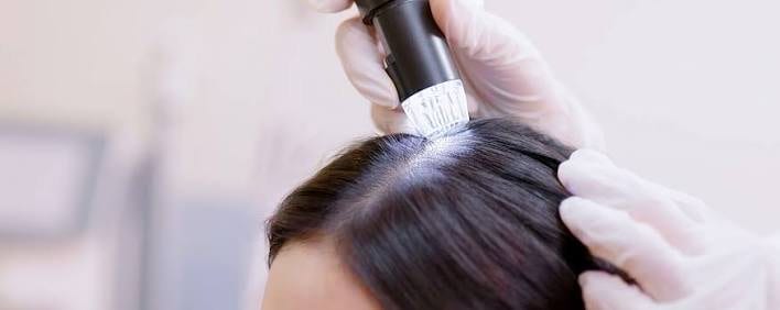
Trichoscopy in donor evaluation
Trichoscopy is magnified examination of the scalp, and it is the tool that turns a donor assessment from an impression into a finding.
Trichoscopy is magnified examination of the scalp, and it is the tool that turns a donor assessment from an impression into a finding. It shows miniaturisation, shaft diameter diversity and unit composition that the naked eye cannot resolve.
What is trichoscopy?
Trichoscopy is dermoscopy applied to the scalp and hair: examination with a handheld or digital magnifying device, usually under polarised light and often with a fixed field of view so counts can be standardised. It is inexpensive, costing less than most clinic furniture, and adds only a few minutes to a consultation.
What can trichoscopy show that the naked eye cannot see?
Trichoscopy reveals shaft diameter diversity (the signature of miniaturisation), the proportion of thin and vellus hairs, hairs per follicular unit, follicular unit density counted within a known field, peripilar signs linked to inflammation, and scarring changes showing lost follicular openings — details invisible to the naked eye.
- Shaft diameter diversity. Terminal hairs of clearly different thickness in the same field. This is the trichoscopic signature of androgen-driven miniaturisation and the single most important finding in donor assessment.
- The proportion of thin and vellus hairs. A rising vellus fraction indicates a follicle population in the process of shrinking.
- Hairs per follicular unit. Whether units contain one, two, three or four hairs — the figure that converts graft counts into hair counts.
- Follicular unit density. Counted within a known field rather than estimated.
- Peripilar signs. Perifollicular pigmentation or a brown halo, associated with the inflammatory component of androgenetic alopecia.
- Scarring changes. Loss of follicular openings, which points toward a cicatricial process rather than androgenetic loss.
Why does trichoscopy matter specifically for donor-area decisions?
A donor area can look normally dense while a substantial proportion of follicles are miniaturising, because visible thinning is a late finding. Grafts from a quietly miniaturising zone keep miniaturising after transfer, looking good at twelve months before fading. Xu and colleagues (2026) found occipital sparing is often assumed rather than verified.
An area can look normally dense to the naked eye while containing a substantial proportion of miniaturising follicles, because a fine hair still occupies its position and still catches light. Visible thinning is a late finding.
For the donor area, that gap between appearance and reality is the whole problem. A donor zone that looks fine but is quietly miniaturising will supply grafts that continue to miniaturise after transfer — a result that looks good at twelve months and fades over the following years, with the donor reserve already spent.
Xu and colleagues made the general point directly in 2026, arguing from paired vertex-occipital assessment that occipital sparing is assumed rather than verified and that donor-area involvement may be overlooked. Trichoscopy is how it stops being assumed.
Where on the scalp should trichoscopy be performed?
Trichoscopy should not rely on a single mid-occipital reading, since density and stability vary across the donor zone. A full assessment checks the mid-occipital reference point, the upper occipital area near the crown transition, above and behind the ears, the nape hairline, and the recipient area for comparison.
One reading from the mid-occipital region is not an assessment. Density and stability vary across the donor zone, and the areas that thin first are the periphery.
- Mid-occipital, as the reference point.
- Upper occipital, toward the crown transition, where harvest drift happens.
- Above and behind the ears, where retrograde thinning shows.
- The nape hairline, which is outside the safe zone and frequently affected.
- The recipient area itself, for comparison — the contrast between donor and recipient miniaturisation is informative in its own right.
What does good trichoscopic documentation look like?
Good documentation means trichoscopic findings are recorded as numbers, not just observed, so a clinic can compare your donor area against itself years later — the only way to know if it has changed. Ask whether images are stored, copies available, and measurements numeric; standardised photographs at consistent length and lighting help too.
How can I tell if a clinic actually assessed my donor area?
Ask not whether a clinic owns a trichoscope but whether they used it on you, where, and what they found. 'Your donor area looks good' is an impression; 'density is 62 follicular units per cm² at the mid-occipital region, 54 above the ears, 2.2 hairs per unit' is an assessment you can plan against.
Sources
- Xu Y, et al. Case Report: Paired vertex-occipital assessment reveals donor-area involvement in diffuse unpatterned alopecia. Frontiers in Medicine, 2026;13:1797275. pubmed.ncbi.nlm.nih.gov/41982548
- Maas D, et al. Rethinking the occipital scalp as a control in advanced androgenetic alopecia. Journal of the American Academy of Dermatology, 2026 (ahead of print). pubmed.ncbi.nlm.nih.gov/42398778
- Jimenez F, Ruifernández JM. Distribution of human hair in follicular units. A mathematical model for estimating the donor size in follicular unit transplantation. Dermatologic Surgery, 1999;25(4):294-298. pubmed.ncbi.nlm.nih.gov/10417585
- Gupta AK, Venkataraman M, Talukder M, Bamimore MA. Finasteride for hair loss: a review. Journal of Dermatological Treatment, 2022;33(4):1938-1946. pubmed.ncbi.nlm.nih.gov/34291720
- Romera de Blas C, Vega Díez D, Ricart Vayá JM, Gómez Zubiaur A. Complications in follicular unit excision hair transplantation: current evidence and practical approaches. Frontiers in Medicine, 2026;13:1750989. pubmed.ncbi.nlm.nih.gov/41709896
This article summarises published research and standard clinical practice. It is general educational information, not medical advice, and it does not replace the instructions your own surgical team gives you. Where their guidance differs from anything here, follow theirs.
Get a no-obligation assessment against fixed criteria, directly on WhatsApp.