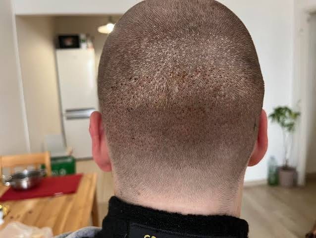
Donor area healing: dot scars and how visible they are
Every FUE extraction leaves a small round scar that usually heals slightly paler than surrounding skin.
Every FUE extraction leaves a small round scar that usually heals slightly paler than surrounding skin. At normal density and moderate hair length they are invisible; visibility depends on punch size, extraction density, skin-hair contrast and how short you wear your hair.
What happens to the skin at each FUE extraction site?
Each punch creates a small circular full-thickness wound that heals by secondary intention, not sutures: a crust forms within days, then contracts and epithelialises over following weeks into a small round scar, usually slightly paler than surrounding skin. This hypopigmentation is a documented complication, and the scars are permanent — FUE is not truly 'scarless'.
Each punch creates a small circular full-thickness wound in the donor scalp. These are left to heal by secondary intention rather than being sutured. Over the first days a tiny crust forms; over the following weeks the wound contracts and epithelialises.
The end result is a small round scar. In most patients it is slightly paler than the surrounding skin, because the healed tissue contains less pigment. Hypopigmentation at donor extraction sites is documented among the recognised donor-area complications of follicular unit excision.
The scars are permanent. This is worth stating plainly, because FUE is sometimes marketed as 'scarless'. It is not scarless; it produces many small scars instead of one linear one.
What is the healing timeline for FUE donor dot scars?
Days 1-3: small pink scabs form and may feel sore. Days 4-10: crusts separate as washing resumes; pinkness persists. Weeks 2-6: redness fades, slower in pale skin. Months 2-4: dots near final appearance and any donor shedding recovers. Months 6-12: final appearance — the only honest point to judge donor-area outcome.
- Days 1-3. Small scabs form over each site. The area is often pink and may feel tight or sore.
- Days 4-10. Crusts separate as washing resumes on the schedule your clinic sets. Pinkness persists.
- Weeks 2-6. Redness fades progressively. Pale-skinned patients typically take longer for the pink to settle.
- Months 2-4. The dots reach close to their final appearance. Any donor shedding that occurred is usually recovering by now.
- Months 6-12. Final appearance. This is the point at which donor-area outcome can honestly be judged — not before.
What determines whether FUE dot scars are ever visible?
Four variables: extraction density (dominant — low density disappears into hair, high density merges into a visibly lighter zone), punch diameter (dot size matches punch size), skin-hair contrast (dark hair on pale skin shows dots earliest), and hair length (invisible above 6mm, visible at a number one or shaved). Below a number three, dots stay hidden.
Four variables account for most of the difference between a donor area nobody notices and one the patient covers up.
- Extraction density. The dominant factor. Dots at low extraction density disappear into surrounding hair; at high density they merge into a visibly lighter zone.
- Punch diameter. Each dot is roughly the size of the punch that made it.
- Skin-hair contrast. Dark hair on pale skin is the highest-contrast combination and shows dots earliest. Low contrast — lighter hair, or darker skin with dark hair — is more forgiving.
- Hair length. At 6 mm and above, most well-executed donor areas are indistinguishable from untouched scalp. At a number one or a shaved head, the dots are visible in any reasonable light.
That last point is the one to internalise before surgery. If you shave your head, you will see the dots. If you never plan to go below about a number three, you probably never will.
What less common donor-area healing complications can occur after FUE?
Beyond ordinary hypopigmentation, documented donor-area complications include hypertrophic scarring (raised, thickened tissue, uncommon, driven mainly by individual healing tendency — disclose any personal history of keloid or hypertrophic scarring at consultation, since it changes risk for FUE and strip harvesting alike), epithelial cysts (usually minor, self-limiting, reported rather than squeezed), and donor depletion.
What options exist to camouflage visible FUE donor scars?
Three options: growing hair longer (simplest, most effective, most common choice); scalp micropigmentation (pigment deposits reducing contrast between pale dots and skin, the most reliable fix, works well short); and grafting beard or body hair into the depleted zone, which spends donor supply to fix a donor problem. None removes the scars, only their visibility.
- Longer hair. The simplest and most effective, and the one most patients settle on.
- Scalp micropigmentation. Small pigment deposits reduce the contrast between pale dots and surrounding skin. It is the most reliable intervention for visible donor scarring and works well at short hair lengths.
- Grafting into the donor area. Occasionally used to add density to a depleted zone, usually with beard or body hair. It spends donor supply to fix a donor problem, so it is a limited option.
None of these removes the scars. They change how visible they are, which for most patients is what actually matters.
Sources
- Romera de Blas C, Vega Díez D, Ricart Vayá JM, Gómez Zubiaur A. Complications in follicular unit excision hair transplantation: current evidence and practical approaches. Frontiers in Medicine, 2026;13:1750989. pubmed.ncbi.nlm.nih.gov/41709896
- Rassman WR, Bernstein RM, McClellan R, Jones R, Worton E, Uyttendaele H. Follicular unit extraction: minimally invasive surgery for hair transplantation. Dermatologic Surgery, 2002;28(8):720-728. pubmed.ncbi.nlm.nih.gov/12174065
- Chauhan K, Tandon M, Kumar A, Taneja N, Hamid SAT. A comprehensive review of evolution of advanced follicular unit excision systems. Journal of Cutaneous and Aesthetic Surgery, 2025;18(2):69-77. pubmed.ncbi.nlm.nih.gov/40212421
- Kim J, Ko YU, Yi KH. The condition of hair follicles produced by different punching methods during FUE surgery. Journal of Cosmetic Dermatology, 2024;23(12):4202-4207. pubmed.ncbi.nlm.nih.gov/39152658
This article summarises published research and standard clinical practice. It is general educational information, not medical advice, and it does not replace the instructions your own surgical team gives you. Where their guidance differs from anything here, follow theirs.
Get a no-obligation assessment against fixed criteria, directly on WhatsApp.