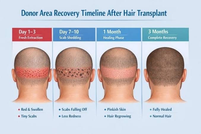
Donor healing timeline week by week
The donor area crusts in the first days, clears over the first week to ten days, loses its redness over the following month, and reaches its final appearance somewhere between six and twelve months.
The donor area crusts in the first days, clears over the first week to ten days, loses its redness over the following month, and reaches its final appearance somewhere between six and twelve months. Judgements about donor outcome before then are premature.
What happens to the donor area in the first three days after a hair transplant?
In the first three days, small crusts form over each site and the area is pink to red, tender and tight, more pronounced after strip surgery around the closure line. Discomfort peaks in this window, making sleep difficult, so sleeping elevated helps both discomfort and swelling. Washing begins on the schedule your clinic sets.
What happens to the donor area between days four and ten after a hair transplant?
Between days four and ten, crusts soften and separate with regular gentle washing, and most patients are clear of crusting by day seven to ten. Pinkness persists, often more noticeable once crusts are gone, while tenderness reduces markedly. This is when the washing protocol matters most — crusts left too long are linked to more folliculitis.
What happens to the donor area during weeks two to six after a hair transplant?
During weeks two to six, redness fades (slower in pale-skinned patients), itching often appears as nerves recover and must not be scratched, folliculitis — small spots as hairs push through healing skin — is common and usually self-limiting, and some patients see donor shock loss, temporary shedding around extraction sites that looks alarming but recovers over months.
Redness fades progressively. Pale-skinned patients typically take longer for the pink to settle, and it can persist for several weeks longer than expected.
Itching frequently appears in this window, associated with healing and with sensory nerves recovering. It can be intense and must not be scratched.
Folliculitis — small spots as hairs push through healing skin — is common from around week two and usually self-limiting.
Some patients experience donor shock loss: temporary shedding of hair around the extraction sites. It looks alarming and it recovers, but it takes months.
What does the donor area look like two to four months after a hair transplant?
By months two to four, redness has largely resolved and extraction sites settle into their final small pale dots. Donor shedding begins recovering on the same schedule as transplanted hair, while numbness keeps improving. The area can look normal at moderate hair length, but patients who assume it is finished at this point are wrong.
When does the donor area reach its final appearance after a hair transplant?
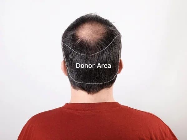
The donor area reaches its true final appearance between months six and twelve — the only honest point to judge donor outcome. By then, density has settled including recovery from shedding, dot scars have reached final contrast, and any hypertrophic scarring or hypopigmentation is apparent, since judging overharvesting earlier is premature either way.
This is when the donor area reaches its true final appearance, and it is the only honest point at which donor outcome can be judged.
- Density has settled, including recovery from any temporary shedding.
- Dot scars have reached their final contrast against surrounding skin.
- Any hypertrophic scarring or persistent hypopigmentation is apparent.
- The hair length at which the donor area still looks untouched can be determined.
The practical implication: do not conclude that a donor area was overharvested at three months, and do not conclude that it was fine either. Both judgements need twelve months.
What should I photograph to track donor area healing after a hair transplant?
Track donor healing by photographing from behind at a known hair length and consistent lighting before surgery, then repeating the shot at three, six and twelve months, and noting your measured pre-operative density and planned extraction proportion. This costs nothing and is the only objective record, since memory is not reliable.
- A pre-operative photograph from behind, at a known hair length and consistent lighting.
- The same photograph at three, six and twelve months.
- A note of your measured pre-operative density and the planned extraction proportion.
This costs nothing and is the only way to have an objective answer later. Memory of what the back of your own head looked like is not reliable, and it is the part of the result you cannot see.
The recipient side of the same weeks is covered under aftercare.
Sources
- Romera de Blas C, Vega Díez D, Ricart Vayá JM, Gómez Zubiaur A. Complications in follicular unit excision hair transplantation: current evidence and practical approaches. Frontiers in Medicine, 2026;13:1750989. pubmed.ncbi.nlm.nih.gov/41709896
- Rassman WR, Bernstein RM, McClellan R, Jones R, Worton E, Uyttendaele H. Follicular unit extraction: minimally invasive surgery for hair transplantation. Dermatologic Surgery, 2002;28(8):720-728. pubmed.ncbi.nlm.nih.gov/12174065
- Bernstein RM, Rassman WR. Graft Anchoring in Hair Transplantation. Dermatologic Surgery, 2006;32(2):198-204. pubmed.ncbi.nlm.nih.gov/16442039
- Giardiello F, De Medeiros Quirino L, Brigante R, Chumak M. Hyperbaric Oxygen Therapy for Enhanced Postoperative Recovery in Hair Transplantation. Cureus, 2025;17(12):e99635. doi.org/10.7759/cureus.99635
- Parsley WM, Perez-Meza D. Review of factors affecting the growth and survival of follicular grafts. Journal of Cutaneous and Aesthetic Surgery, 2010;3(2). jcasonline.com
This article summarises published research and standard clinical practice. It is general educational information, not medical advice, and it does not replace the instructions your own surgical team gives you. Where their guidance differs from anything here, follow theirs.
Get a no-obligation assessment against fixed criteria, directly on WhatsApp.